


60-year-old mystery solved: Molecular model of heart discovered by Bengali scientist – GetBengal story

Say it with your heart and not with your head! Often said but not always followed, for even today despite a big leap in medical science, everything about the heart is still not written, and many mysteries are still not solved. The heart is one of the most complex and arguably the most vital organs within the human body. Although most can say that the heart pumps blood (a given fact about the human anatomy – a concept depended upon for its faithful delivery), the human heart proves to be an extremely complex and necessary organ of the human body. It beats 100,000 times per day while pumping 5 gallons of blood each minute through the 60,000 miles worth of vessels in the body – this is about 2,000 gallons of blood pumped per day and supports the body’s daily demand.
For the past 60 years, scientists across the globe have been researching the molecular formation of the heart. Recently, a team of international researchers that included in the forefront a Bengali scientist, has cracked the puzzle and has been able to map out an important part of the heart on a molecular level. The study titled ‘Cryo-EM structure of the human cardiac myosin filament’ was published in ‘Nature.’ The team comprising Debabrata Dutta, Ph.D.; Raúl Padrón, Ph.D.; and Roger Craig, Ph.D., authored this work, which answers questions about heart muscle structure, physiology and disease that have remained a mystery for the last 60 years.
Dutta has throughout been a brilliant student and he always dreamt of being a scientist from a very early age. Hailing from Jungalmahal, once a Maoist hotbed and now battling hunger and anger, the vast underdeveloped hinterland has very little to offer for its prodigious children. Dutta however was an exception. He studied at Bankura Christian Collegiate School and after completing his Higher Secondary from there, he shifted to Kolkata to pursue his graduation from St. Xavier’s College with honours in Chemistry. He obtained his Master’s degree from the University of Calcutta and then moved to the Indian Institute of Technology (IIT), Kharagpur to do research on biotechnology. Dutta spread his wings in the country’s premier research institution, recognized as an Institute of national Importance. He completed his PhD in 2019 and then flew to the US for higher studies. There he joined the American National Institute of Health and then moved on to the University of Massachusetts Medical School (UMass Chan Medical School) to continue his research in Dr. Roger Craig’s laboratory in the Radiology Department.
“From here began my seminal work on mapping out an important part of the heart on a molecular level. The pioneering work resolves past uncertainties and integrates previous data on cardiac muscle structure and function. It provides a new paradigm for interpreting structural, physiological and clinical observations, and for the design of potential therapeutic drugs,” says Dutta.

Dutta explains that we are more or less familiar with heart sounds generated by the beating heart and the resultant flow of blood through it. In healthy adults, there are two normal heart sounds, often described as a lub and a dub that occur in sequence with each heartbeat. These are the first heart sound (S1) and second heart sound (S2), produced by the closing of the atrio-ventricular valves and semilunar valves, respectively. In addition to these normal sounds, a variety of other sounds may be present including heart murmurs, adventitious sounds, and gallop rhythms S3 and S4.
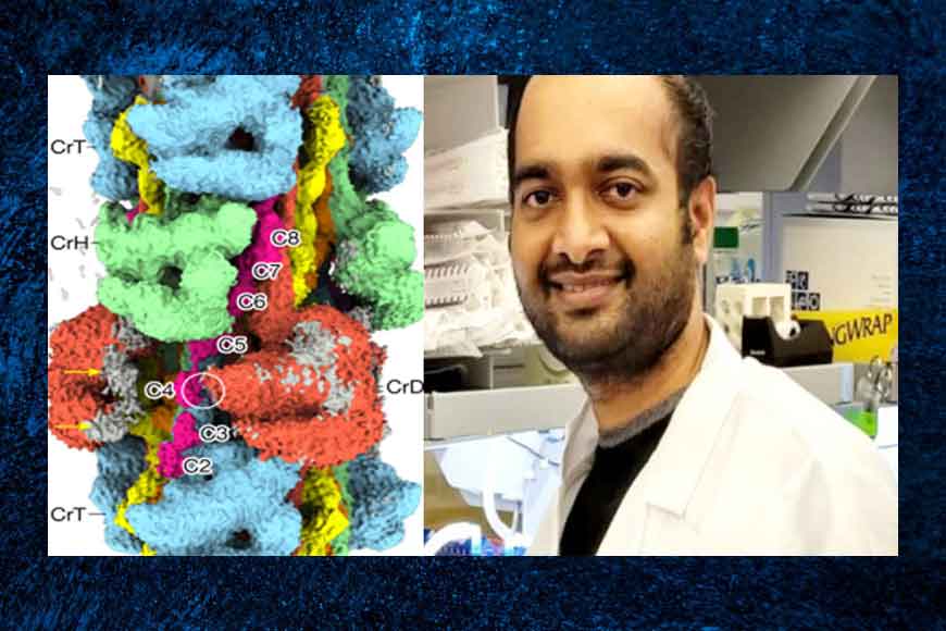
The heart is made up of billions of cells. Each cell contains thousands of smaller structures, called sarcomeres. These are the building blocks of muscle. Within each block are hundreds of myosin filaments. Each filament has roughly 2,000 molecules arranged in a complicated structure that scientists have been trying to understand for decades. They knew quite a lot about the individual molecules, and people thought the myosins could be arranged in groups of six that were called crowns, but not much beyond that. However, the most interesting discovery in the paper is that there are three different types of crowns.
The pumping of the heart is powered by filaments of the motor protein myosin that pull on actin filaments to generate cardiac contraction. In addition to myosin, the filaments contain cardiac myosin-binding protein C (cMyBP-C), which modulates contractility in response to physiological stimuli, and titin, which functions as a scaffold for filament assembly. Myosin, cMyBP-C and titin are all subject to mutation, which can lead to heart failure. Despite the central importance of cardiac myosin filaments to life, their molecular structure has remained a mystery for 60 years.
With the new discovery, it becomes clear that heart muscle can be controlled more precisely than we thought. The new discovery also revealed how myosin binding protein-C, another protein that is linked to genetic heart disease, sits within the structure. It provides scientists a new level of information about how the molecules are arranged in the heart. Dr. Dutta and his teammates solved the structure of the main (cMyBP-C-containing) region of the human cardiac filament using cryo-electron microscopy. The research group produced single-particle 3D reconstructions of the cardiac thick filaments. The pictures provide a new framework for interpreting structural, physiological and clinical observations. The reconstruction reveals the architecture of titin and cMyBP-C. It shows how myosin’s motor domains (heads) form three different types of motif (providing functional flexibility), which interact with each other and with titin and cMyBP-C to dictate filament architecture and function. The packing of myosin tails in the filament backbone is also resolved. The structure suggests how cMyBP-C helps to generate the cardiac super-relaxed state; how titin and cMyBP-C may contribute to length-dependent activation; and how mutations in myosin and cMyBP-C might disturb interactions, causing disease. The reconstruction resolves past uncertainties and integrates previous data on cardiac muscle structure and function. It provides a new paradigm for interpreting structural, physiological and clinical observations, and for the design of potential therapeutic drugs.
Cardiovascular diseases (CVD) are the leading cause of death and disability in India. The CVD epidemic in Indians is characterized by a higher relative risk burden, an earlier age of onset, higher case fatality and higher premature deaths. For decades, researchers have been trying to understand the reason for this increased burden and propensity of CVD among Indians. Following the World Health Organization, India accounts for one-fifth of these deaths worldwide, especially in the younger population. The results of the Global Burden of Disease study state the age-standardized Cardiovascular Disease (CVD) death rate of 272 per 100000 population in India which is much higher than that of the global average of 235. “Our study gives a much better understanding of how the molecules in our hearts interact and thus will largely help doctors to understand the working of the heart muscles and how they can malfunction or can be fixed.”


